SCANNING ELECTRON
MICROSCOPE
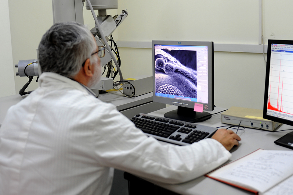
The Scanning Electron Microscope (SEM) is a tool that enables ultra-high-resolution observation of surface morphology at the sub micrometric scale, with a resolution down to just a few tens of nanometers.
The SEM works by directing a focused beam of high-energy electrons onto the sample surface. As these electrons interact with the atoms in the specimen, they produce a variety of signals that can be detected and transformed into highly detailed images. These images provide crucial information about the sample’s surface structure, texture, and topography.
In addition to imaging, our SEM system is equipped with Energy Dispersive X-ray Spectroscopy (EDS), which allows us to perform elemental analysis. This technique identifies and quantifies the chemical elements present in a selected area of the sample, offering essential insights into its composition.